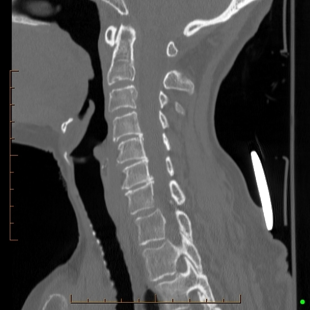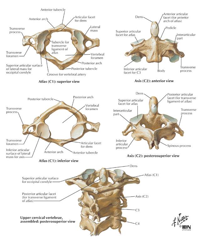
CT is necessary for complete assessment of these injuries, especially in young children, as atlas may not be visualized on radiographs. Retropharyngeal space width of more than 5 mm at the level of C3 body is the indirect sign of anterior arch fracture on lateral radiographs. Vertical anterior arch fracture or a “plough fracture” can be occult on radiography, and the comminuted, anteriorly displaced bone fragments are best seen on axial CT images. Horizontal fracture of the anterior arch is detected on lateral radiograph as a lucent, irregular, noncorticated line and is best appreciated on sagittal CT images. It occurs most often near the junction with lateral masses, and is best appreciated on axial CT images due to its most common vertical orientation. Fracture of the posterior arch is seen on lateral radiographs as a lucent, noncorticated line through the posterior element. These injuries are classified as Type I fractures of the atlas.

Stable fractures can be treated with rigid cervical collar for 8–12 weeks, while comminuted or unstable fractures are treated either with a rigid cervical orthosis or a halo vest. Furthermore, MR is crucial in assessing possible spinal cord injuries by dislocated bone fragments, and is indicated in patients with neurologic deficit. Transverse ligament rupture can be well appreciated on axial T1 and T2 MR images as hypointense ligament band disruption. Assessed on CT imaging, avulsion at the transverse ligament insertion at the medial part of C1 lateral mass and comminuted lateral mass fracture indicate Dickman type II transverse ligament lesion. Atlanto-axial lateral displacement greater than 7 mm and atlanto-dental interval greater than 3 mm are indirect signs of transverse ligament rupture recognized on open-mouth and lateral radiographs. Integrity of the transverse ligament determines the stability of C1 lateral mass fracture and guides further imaging and treatment. Conservative management consists of rigid collar placement with 6-week follow-up dynamic radiographs. Surgical treatment is indicated for patients with AO offset (>2 mm) or evidence of significant neural compression and may be with occipito-cervical fusion or halo placement. focused on 415 reported cases and concluded that nearly all occipital condyle fractures can be managed nonsurgically. The 2013 meta-analysis by Theodore et al. on 100 patients (25% of whom were lost to follow-up) treated patients with occipital condyle fractures and less than 2 mm of atlanto-occipital (AO) joint offset conservatively with good outcomes. Criteria for the stability of occipital condyle fractures are not universally defined.

Lower cranial nerve palsies are present in one-third of patients and may present in a delayed fashion. More important is careful inspection of CT images for additional injuries of the skull, atlas, and axis because of the significant forces involved and anatomic proximity of critical structures.Ĭlinical Findings, Implications, and Treatmentīecause of the associated high-energy mechanism of injury, patients with occipital condyle fractures frequently are obtunded at presentation otherwise, pain is the predominant complaint. The role of MRI for direct inspection of the alar ligaments and tectorial membrane is questionable. Anderson classification is more widely accepted. This group suggested that more than 8 degrees of rotation or 1 mm of translation of the occiput relative to C1, direct evidence of alar ligament avulsion, or MRI evidence of atlanto-axial disruption is consistent with instability.

proposed a classification scheme consisting of type 1 (nondisplaced), type 2a (displaced stable), and type 2b (displaced unstable) fractures. These fractures are classified into three types by the Anderson and Montesano scheme: Type I – comminuted and nondisplaced secondary to axial loading Type II – extend into the condyle from a linear fracture in the remainder of the skull base (generally involve the base of the occipital condyle without complete separation of the condyle from the skull) Type III – avulsion fractures of the occipital condyle.

Occipital condyle fractures need to be evaluated with multiplanar CT, as they may at times be clearly seen in a single plane only. 19 Isolated Fracture of the Anterior or Posterior Arch of AtlasĢ5 Vertebral Body Microfractures / Bone Marrow EdemaĢ6 Jefferson Burst Fractures of the AtlasĢ7 Burst Fractures (Other Than Jefferson Fracture)ĭaniela Distefano and Alessandro Cianfoni


 0 kommentar(er)
